- Research Services
- Capabilities
- About Us
- Resources
- Contact Us
 Tissue and Sample Processing
 Cellular and Molecular Characterization
 Gene Expression Analysis
 Blood/Sample Analysis
 In Vitro and Ex Vivo Capabilities
In vitro and ex vivo analyses are important components of a complete preclinical program. Whether intended to complement an existing in vivo project or indicated as standalone experiments, BioModels has the tools and expertise to generate data that supports therapeutic efficacy and mechanism of action studies.
BioModels offers a fully equipped in vitro facility to support:
Complete your preclinical program with BioModels’ comprehensive tissue and sample processing capabilities. We offer established and custom approaches to characterize disease phenotypes, evaluate target engagement and mechanism of action, whether in support of validated animal models or as standalone experiments. Processing and analysis of samples generated in your lab or by collaborators is also available.
Support your preclinical program with BioModels’ comprehensive cellular and molecular analysis capabilities. We offer established and custom approaches to characterize disease phenotypes and evaluate target engagement and mechanism of action, whether in support of validated animal models or as standalone experiments. Analysis of samples generated in your lab or by collaborators is also available.
Support your preclinical program with BioModels’ gene expression analysis capabilities. We offer established and custom approaches to characterize disease phenotypes and evaluate target engagement and mechanism of action, whether in support of validated animal models or as standalone experiments. Analysis of samples generated in your lab or by collaborators is also available.
Support your preclinical program with BioModels’ comprehensive blood/sample analysis capabilities. We offer established and custom approaches to characterize disease phenotypes and evaluate target engagement and mechanism of action, whether in support of validated animal models or as standalone experiments. Analysis of samples generated in your lab or by collaborators is also available.
Capabilities

Tissue homogenization and pulverization is essential for generating tissue lysates and supernatants that can be used for a variety of downstream applications. BioModels has experience with mechanical high-throughput homogenization and pulverization of a wide range of tissues and organs. Custom protocol development is also available to accommodate your unique endpoints.

Close

Tissue dissociation is essential for generating single-cell suspensions that can then be used for a variety of downstream applications. BioModels has experience with dissociation of many different tissues and organs with semi-automated options for increased throughput, and standardized systems to ensure reproducibility. Biomodels protocols for tissue dissociation are also flexible, allowing for customization that accommodates your unique applications.
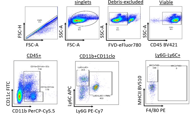
Spleens from DSS-administered animals are subject to dissociation with a gentleMACS™ Dissociator, and a splenocyte single-cell suspension is produced. Splenocytes are stained with a panel of antibodies for identification of dendritic cells, neutrophils, macrophages, and monocytes.

Close

Complex tissue digestion and cell sorting techniques enable exciting downstream analyses that depend on obtaining a quality sample. Isolating specific cell types from complex organ matrixes can be challenging, and the BioModels team is here to help. We offer a number of off-the-shelf digestion and cell sorting protocols designed to enrich for specific cell types from a variety of organs, including enrichment for immune cells from the intestinal lamina propria, lung, and more. Custom protocol development options are also available.
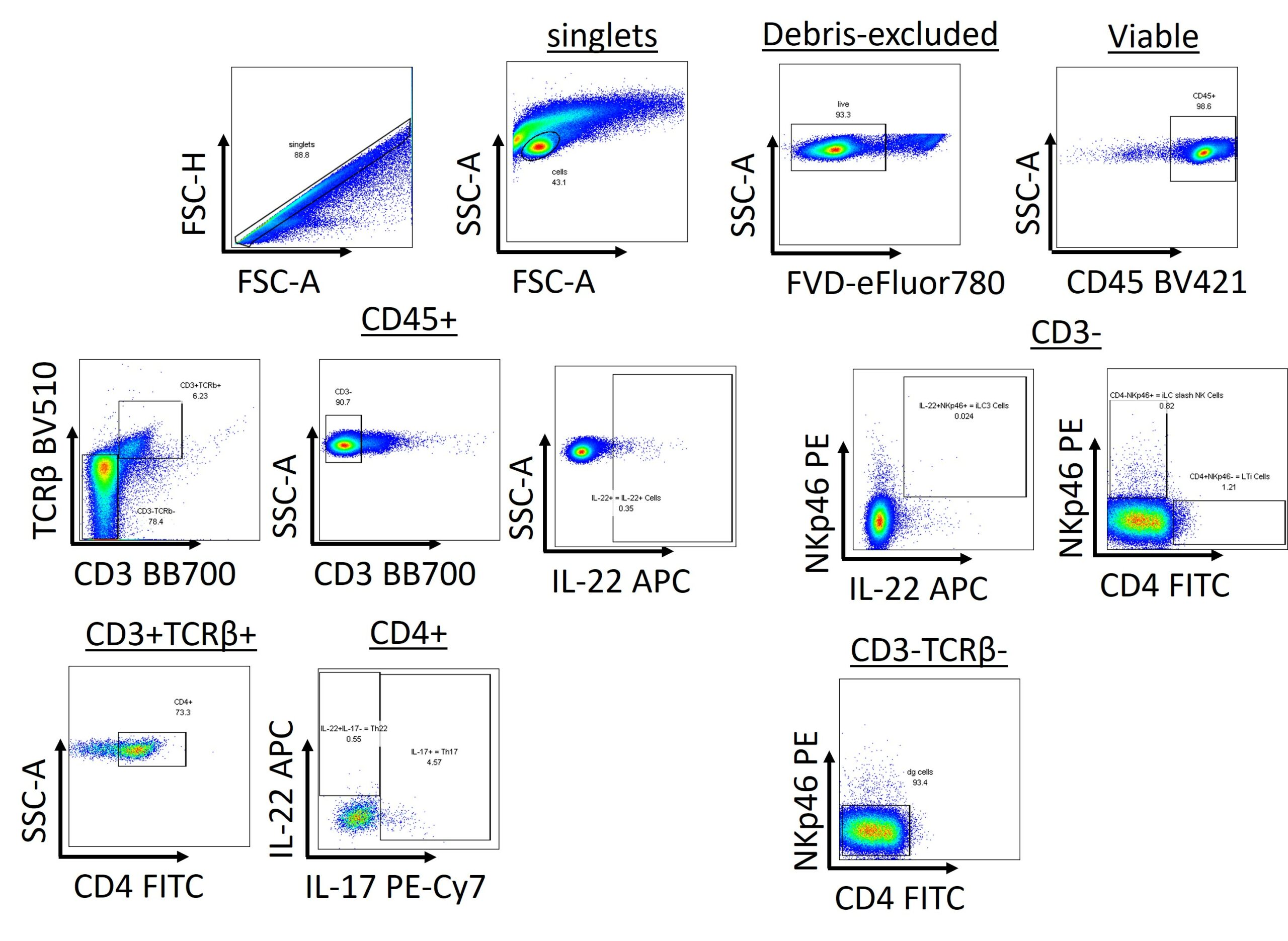
Intestines from DSS-administered animals undergo a series of enzymatic digestions to enrich for lymphocytes. Lymphocytes are stimulated with PMA/Ionomycin and subsequently stained with a panel of antibodies for identification of αβ T cells, TH cells, TH17 cells, TH22 cells, LTi cells, iLC/NK cells, and iLC3 cells.

Close

Histopathology and immunohistochemistry are important capabilities with applicability throughout the drug discovery pipeline. We offer access to this critical service through our collaboration with the experts at Dallas Tissue Research (DTR). The team at DTR provides non-clinical, non-GLP histology, advanced staining, slide scanning, image analysis, and pathology services

Close
Capabilities

Flow cytometry is a powerful and widely published tool, with utility across the drug discovery pipeline. Routinely used for immunophenotyping, flow cytometry enables comprehensive analysis of immune cell subsets, activation status, and cytokine production from a variety of sample types. BioModels’ numerous pre-validated panels enable off-the-shelf immune cell characterization. Custom panel design options support experiments unique to your program and hypothesis.
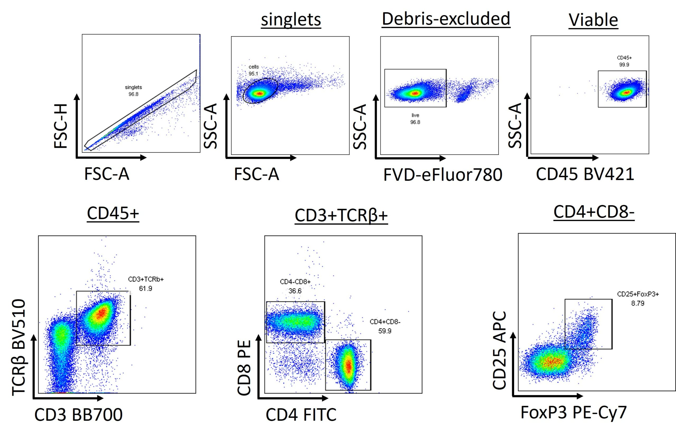
mLN are processed to single cell suspension and stained with a panel of antibodies for identification of αβ T cells, TH cells, Tc cells, and Tregulatory cells

Close

Multiplex cytokine analysis allows you to simultaneously detect levels of multiple cytokine and chemokine biomarkers in a single sample, and thereby ensuring you get the most data out of your samples. Multiplex cytokine analysis is useful for identifying biomarkers distinguishing healthy and disease states in a variety of sample types. BioModels offers options for pre-validated panels which enable cytokine characterization for off-the-shelf disease models. Custom panel options to support experiments unique to your program and hypothesis are also available
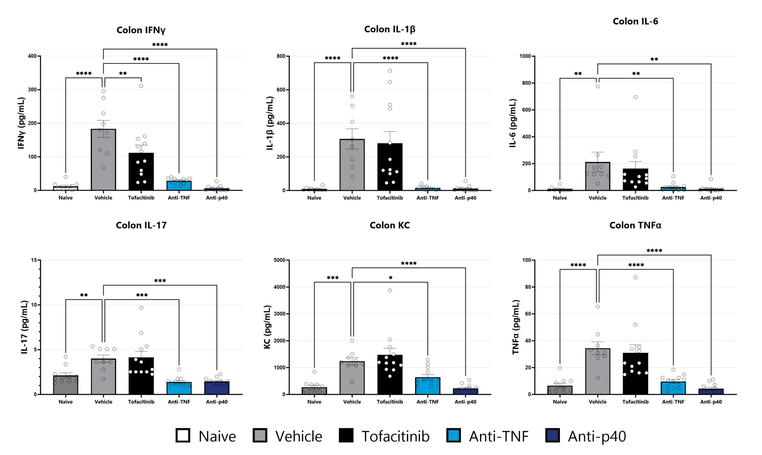
Colon samples from the CD40 mAb-induced colitis are homogenized and assessed via multiplex cytokine analysis for IFNγ, IL-1β, IL-6, IL-17, KC, and TNFα. Additional cytokines are available. (*p<0.05; **p<0.01; ***p<0.005; ****p<0.001 compared to the vehicle-control).

Close

Bronchoalveolar lavage (BAL) is an experimental procedure that allows the investigator to examine the cellular and acellular contents of the lung lumen in healthy and diseased animals. Cellular contents from the lung can be identified either by staining cells with Diff-Quik or by flow cytometry to distinguish macrophages, neutrophils, eosinophils, and lymphocytes. Acellular contents can be analyzed for specific cytokines/soluble analytes and total protein content.
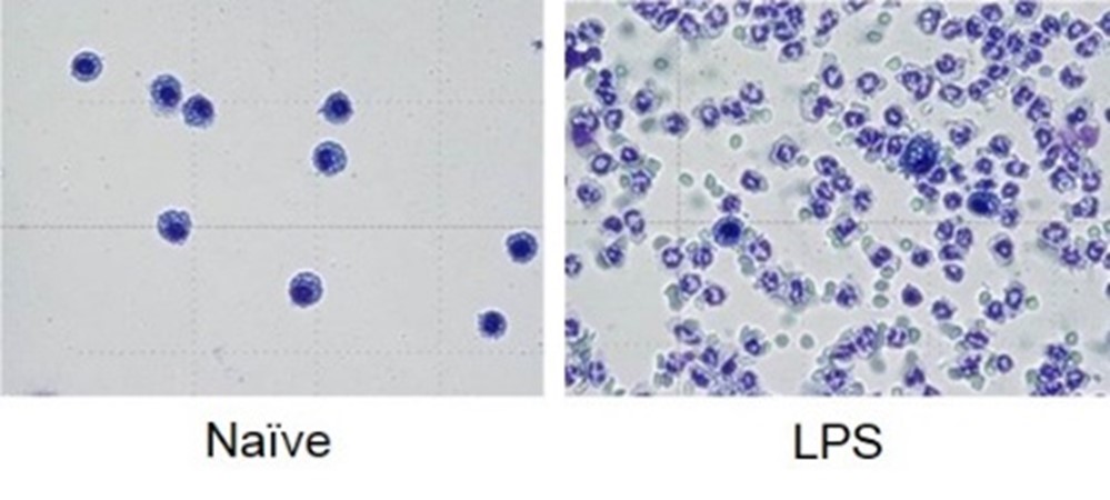
BALB/c mice are administered LPS on Day 0 and BAL fluid is assessed at 24 hours following Diff-Quik staining
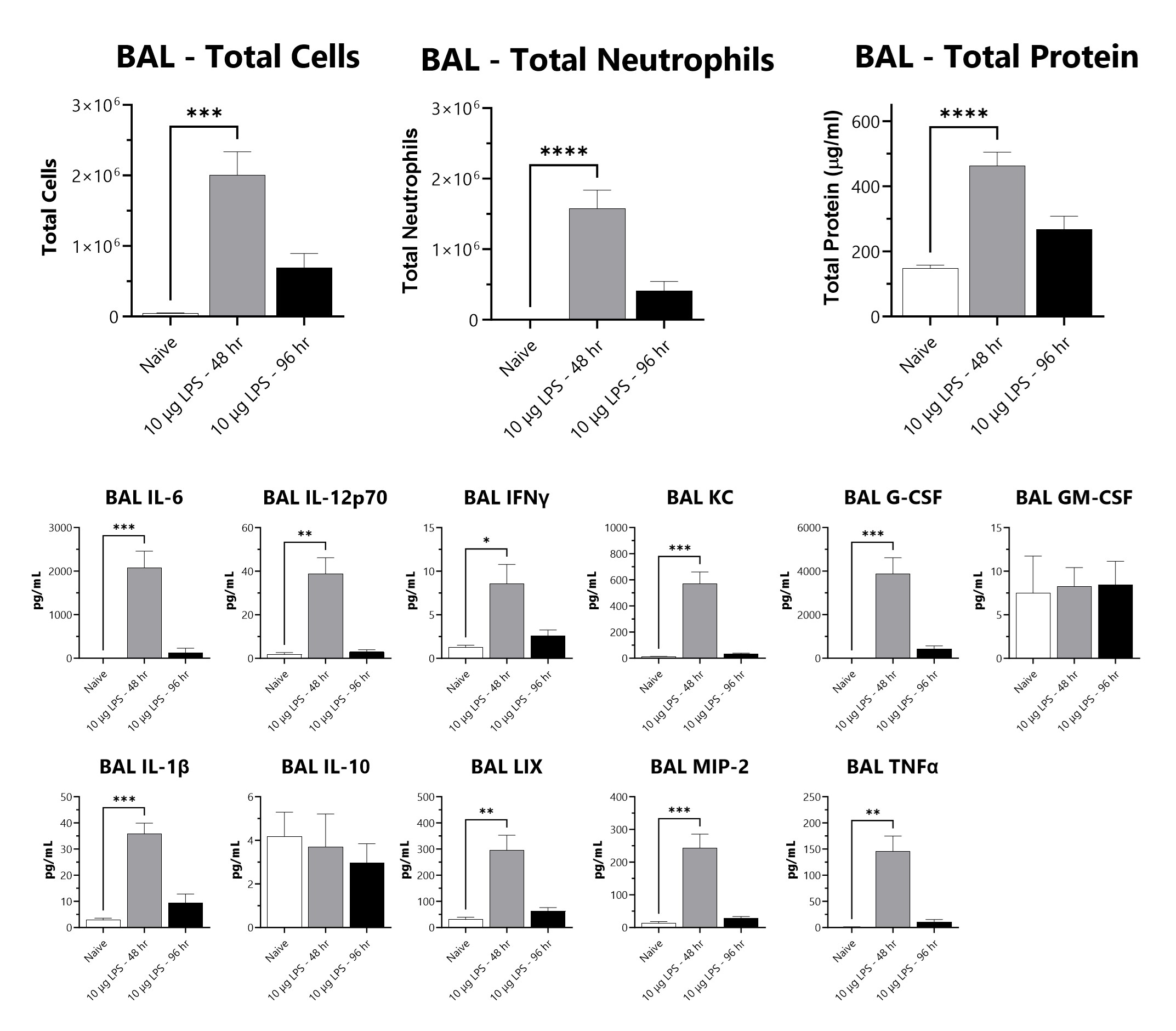
BALB/c mice are administered LPS on Day 0 and BAL fluid is assessed at 48 or 96 hours for total cells, total neutrophils, total protein, and cytokines following administration 10 µg of LPS. (*p<0.05; **p<0.01; ***p<0.001; ****p<0.0001 compared to the naïve control)

Close

ELISA and other plate-based assays allow for detection and quantification of specific analytes in drug discovery and development. These assays allow you to quantitate levels of single cytokines, secreted proteins, and intracellular proteins from tissue lysates in a wide range of sample types. BioModels’ pre-validated assays enable disease characterization of a number of established disease-specific biomarkers as well as custom targets. Custom assays and assay development are also an option to support experiments unique to your program and hypotheses.
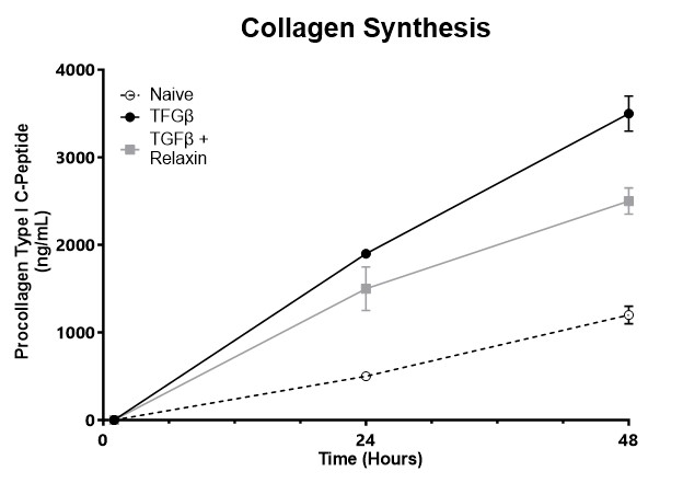
Pro-collagen Type 1 C-Peptide (PIP) is measured in cell culture supernatant of normal human lung fibroblasts by ELISA. Data represents the amount of PIP secreted by cells as an indicator of the development of fibrosis.

Close
Capabilities

Nucleic acid (RNA and DNA) purification is an initial and crucial step in many molecular biology and genomic workflows. The need for high-quality, highly pure nucleic acid is important for a wide range of research applications including quantification of mRNA by qRT-PCR. BioModels’ optimized RNA/DNA isolation protocols for a variety of sample types enables high-quality RNA/DNA yields. Custom isolation protocol options to support experiments unique to your program and hypothesis are also availabl

Close

Reverse transcription polymerase chain reaction (RT-PCR) is a widely used tool in genomic workflows. RT-PCR allows for the detection of low abundance RNAs in a sample, and production of the corresponding cDNA, thereby facilitating the cloning of low copy genes. BioModels’ optimized RT-PCR protocols for a variety of sample types enables high-throughput cDNA synthesis. Custom RT-PCR protocol options to support experiments unique to your program and hypothesis are also available.

Close

Quantitative PCR (qPCR) or quantitative RT-PCR (qRT-PCR) is among the most valuable tools in biological research. qPCR/qRT-PCR enables rapid and sensitive determination and quantitation of nucleic acid in various biological samples, with diverse applications such as gene expression analysis. BioModels’ numerous pre-validated genes enable off-the-shelf disease characterization. Custom qPCR/qRT-PCR protocol options to support experiments unique to your program and hypothesis are also available.
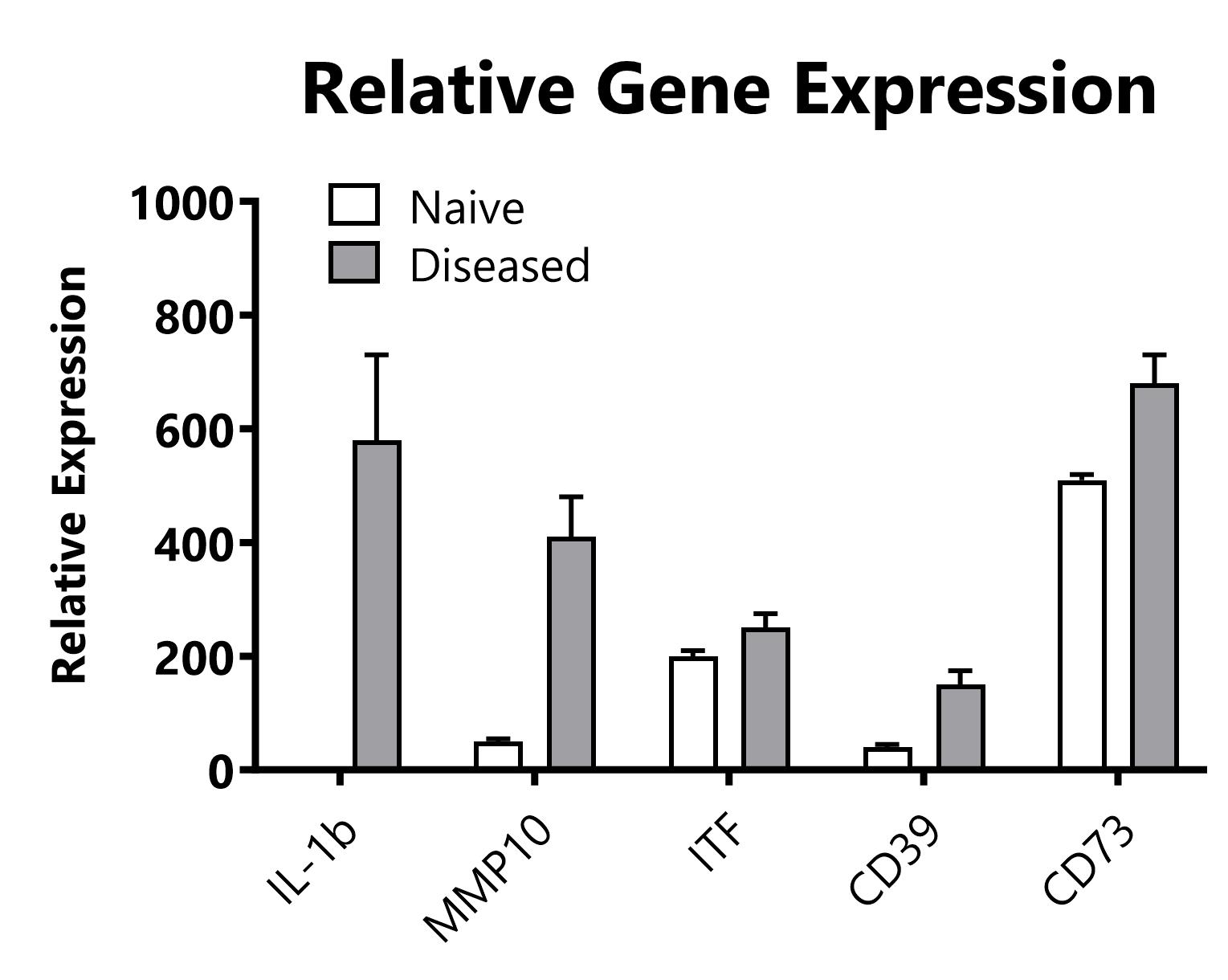
RNA is isolated from colon tissue, and processed to cDNA which is then analyzed by qPCR to assess relative gene expression levels of inflammatory genes

Close
Capabilities

Blood chemistry analysis allows for longitudinal assessment of multiple systemic markers of disease in a single sample. Using a small 100 µL serum or plasma sample, quantitative values are reported for the specified readouts providing a snapshot of the animal’s health or a downstream effect of an experimental treatment. Analyze blood samples generated at BioModels in one of our preclinical animal models, or assess samples generated in your lab or by a collaborator.
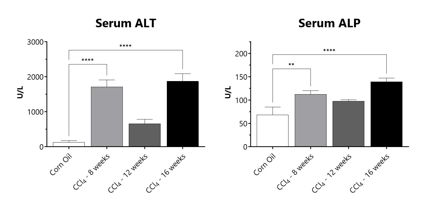
Serum samples from CCl4 administered animals are assessed for levels of alanine transaminase (ALT) and alkaline phosphatase (ALP). (**p<0.01; ****p<0.0001 compared to the corn oil control group)

Close

Complete blood count analysis allows for longitudinal assessment of leukocyte subsets, as well as other blood components such as hemoglobin or platelets, that may be of interest for your study or program. Using a small 50 µL blood sample, quantitative values are reported for the specified readouts providing a snapshot of the animal’s health or a downstream effect of an experimental treatment. Analyze blood samples generated at BioModels in one of our preclinical animal models, or assess samples generated in your lab or by a collaborator.

Close

Blood glucose monitoring allows for longitudinal assessment of the metabolic state of animals throughout a study. Using a single drop of blood, quantitative glucose levels can be assessed in animals. Analyze blood samples generated at BioModels in one of our preclinical animal models.
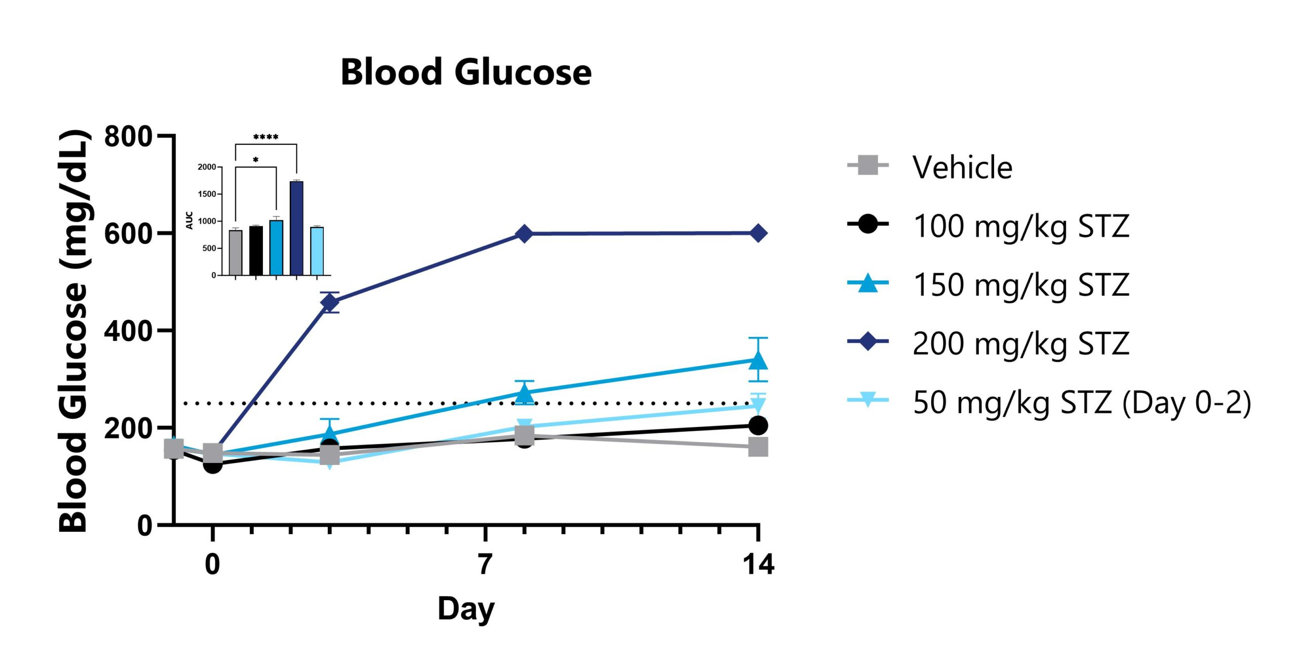
Blood glucose levels are monitored using a blood glucose reader. The AUC is calculated to compare groups and is shown in the inset. (*p<0.05; ****p<0.0001).

Close

Flow cytometry is a powerful and widely published tool, with utility across the drug discovery pipeline. Routinely used for immunophenotyping, flow cytometry enables comprehensive analysis of immune cell subsets, activation status, and cytokine production from a variety of sample types. BioModels’ numerous pre-validated panels enable off-the-shelf immune cell characterization. Custom panel design options support experiments unique to your program and hypothesis.
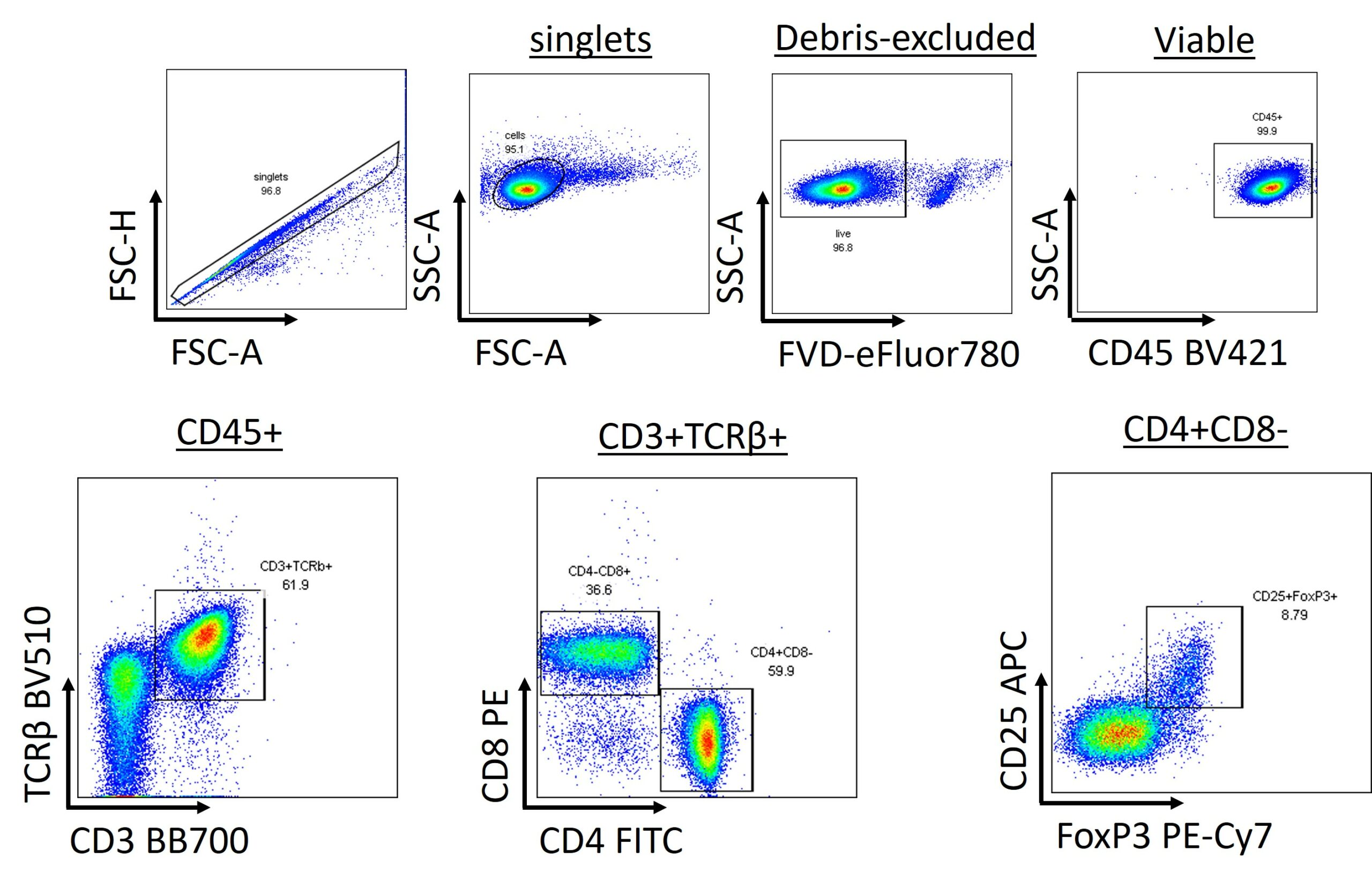
mLN are processed to single cell suspension and stained with a panel of antibodies for identification of αβ T cells, TH cells, Tc cells, and Tregulatory cells.

Close

Bronchoalveolar lavage (BAL) is an experimental procedure that allows the investigator to examine the cellular and acellular contents of the lung lumen in healthy and diseased animals. Cellular contents from the lung can be identified either by staining cells with Diff-Quik to distinguish macrophages, neutrophils, eosinophils, and lymphocytes.
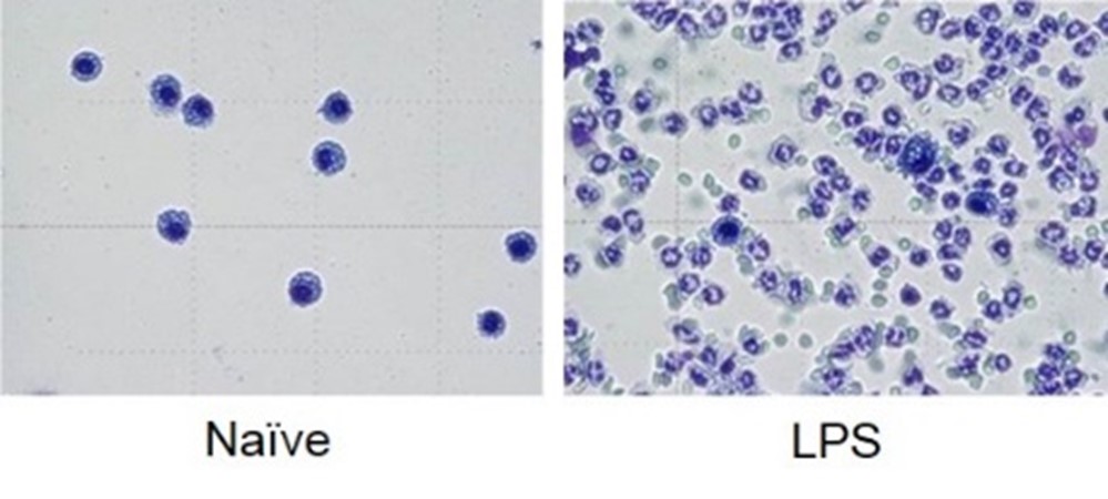
BALB/c mice are administered LPS on Day 0 and BAL fluid is assessed at 24 hours following Diff-Quik staining.
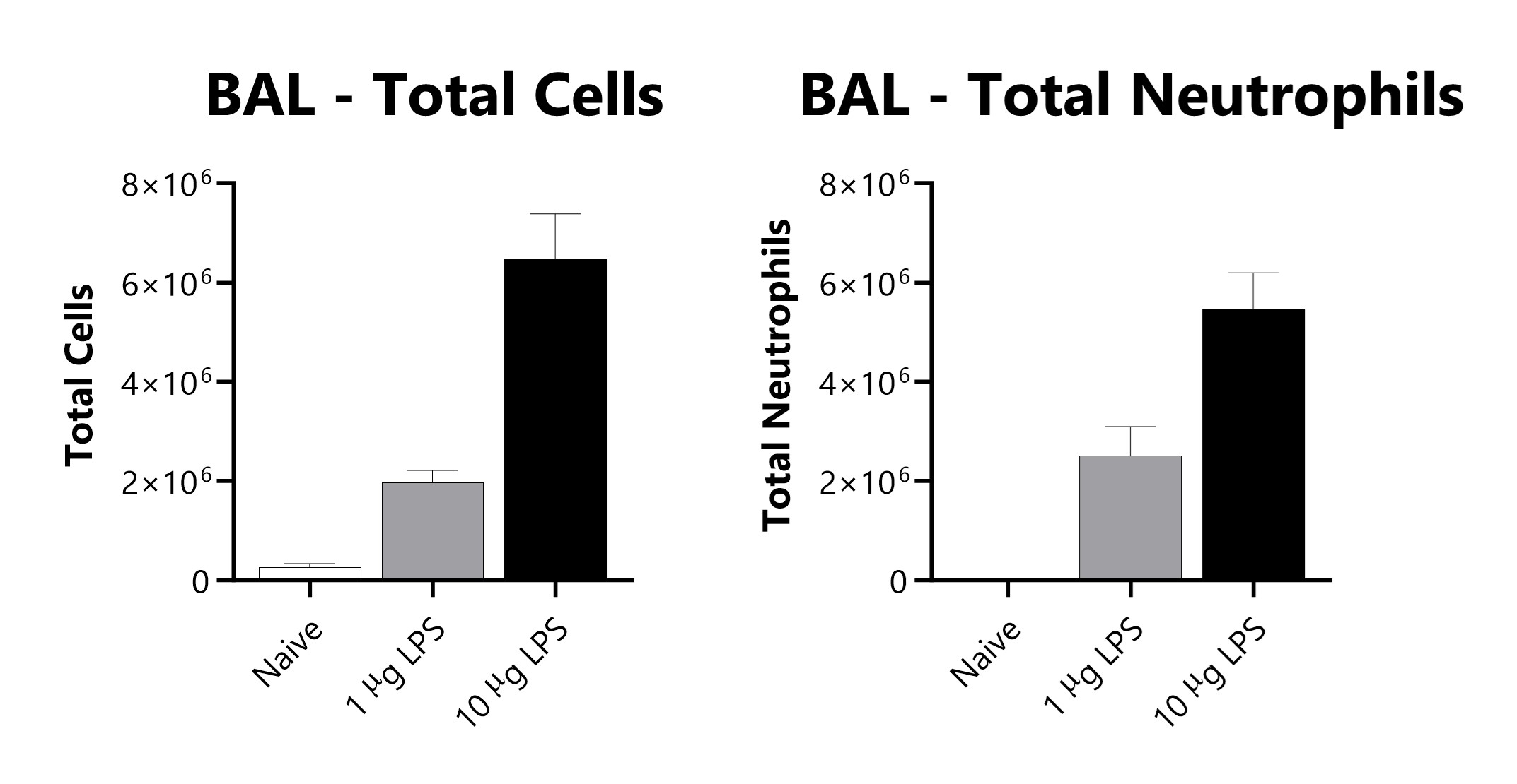
BALB/c mice are administered LPS on Day 0 and BAL fluid is assessed at 48 or 96 hours for total cells and total neutrophils following administration 10 µg of LPS. ***p<0.001; ****p<0.0001 compared to the naïve control).

Close


